

How To Draw Nmr Spectra From Structure
NMR - Interpretation
- Page ID
- 1812
Nuclear Magnetic Resonance (NMR) interpretation plays a pivotal role in molecular identifications. As interpreting NMR spectra, the structure of an unknown compound, every bit well every bit known structures, can be assigned by several factors such as chemical shift, spin multiplicity, coupling constants, and integration. This Module focuses on the nigh of import 1H and 13C NMR spectra to find out structure fifty-fifty though there are diverse kinds of NMR spectra such every bit xivDue north, xixF, and 31P. NMR spectrum shows that x- axis is chemical shift in ppm. Information technology also contains integral areas, splitting pattern, and coupling constant.
Strategy for Solving Construction
Hither is the general strategy for solving structure with NMR:
- Molecular formula is determined by chemical analysis such every bit simple analysis
- Double-bail equivalent (also known as Degree of Unsaturation) is calculated by a simple equation to estimate the number of the multiple bonds and rings. It assumes that oxygen (O) and sulfur (S) are ignored and element of group vii (Cl, Br) and nitrogen is replaced by CH. The resulting empirical formula is CaHb
.jpg?revision=1)
- Structure fragmentation is determined by chemical shift, spin multiplicity, integral (peak expanse), and coupling constants (\(^1J\), \(^2J\))
- Molecular skeleton is built up using 2-dimensional NMR spectroscopy.
- Relative configuration is predicted by coupling constant (3J).
oneH NMR
Chemical Shift
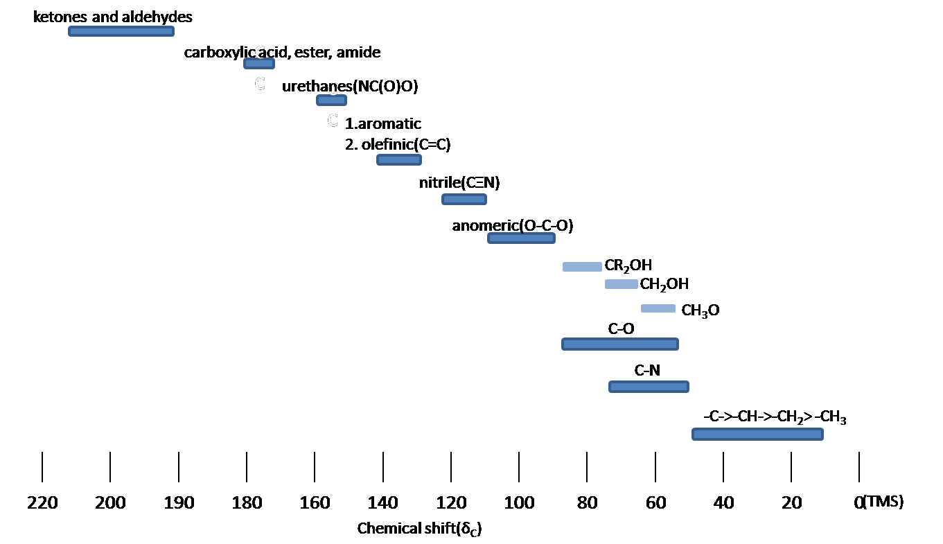
Chemic shift is associated with the Larmor frequency of a nuclear spin to its chemic environment. Tetramethylsilane (TMS, \(\ce{(CH3)4Si}\)) is more often than not used as an internal standard to decide chemical shift of compounds: δTMS=0 ppm. In other words, frequencies for chemicals are measured for a 1H or xiiiC nucleus of a sample from the 1H or 13C resonance of TMS. Information technology is of import to understand tendency of chemical shift in terms of NMR interpretation. The proton NMR chemical shift is affect by nearness to electronegative atoms (O, N, halogen.) and unsaturated groups (C=C,C=O, aromatic). Electronegative groups move to the down field (left; increase in ppm). Unsaturated groups shift to downfield (left) when affecting nucleus is in the aeroplane of the unsaturation, but reverse shift takes place in the regions above and below this plane. aneH chemical shift play a function in identifying many functional groups. Figure \(\PageIndex{1}\). indicates of import example to figure out the functional groups.

Chemical equivalence
Protons with Chemical equivalence has the aforementioned chemical shift due to symmetry within molecule (\(CH_3COCH_3\)) or fast rotation around single bond (-CHthree; methyl groups).
Spin-Spin Splitting
Spin-Spin splitting means that an absorbing pinnacle is split by more than ane "neighbor" proton. Splitting signals are separated to J Hz, where is called the coupling constant. The spitting is a very essential part to obtain verbal data about the number of the neighboring protons. The maximum of altitude for splitting is three bonds. Chemical equivalent protons do not consequence in spin-spin splitting. When a proton splits, the proton's chemic shift is determined in the middle of the splitting lines.
Spin Multiplicity (Splitting pattern)
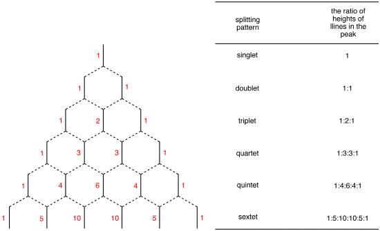
Spin Multiplicity plays a function in determining the number of neighboring protons. Here is a multiplicity rules: In case of \(A_mB_n\) system, the multiplicity rule is that Nuclei of \(B\) element produce a splitting the \(A\) signal into \(nB+1\) lines. The general formula which applies to all nuclei is \(2_nI+1\), where \(I\) is the spin breakthrough number of the coupled chemical element. The relative intensities of the each lines are given past the coefficients of the Pascal's triangle (Figure \(\PageIndex{2}\)).
Kickoff-club splitting blueprint
The chemical shift difference in Hertz between coupled protons in Hertz is much larger than the \(J\) coupling constant:
\[ \dfrac{\Delta \nu }{J} \ge viii\]
Where \(\Delta \nu\) is the deviation of chemical shift. In other give-and-take, the proton is just coupled to other protons that are far away in chemical shift. The spectrum is called first-guild spectrum. The splitting pattern depends on the magnetic field. The 2nd-social club splitting at the lower field can exist resolved into first-order splitting pattern at the high field. The beginning-order splitting pattern is allowed to multiplicity rule (Due north+1) and Pascal's triangle to determine splitting blueprint and intensity distribution.
Example \(\PageIndex{i}\)
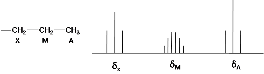
The note is that structure system is AiiiChiliadtwoXtwo. Ha and Hx has the triplet pattern by Hm because of N+1 rule. The point of Hm is split into six peaks past Hx and Ha (Figure3) The First lodge pattern easily is predicted due to separation with equal splitting pattern.
High-guild splitting pattern
High-order splitting pattern takes place when chemical shift deviation in Hertz is much less or the same that society of magnitude as the j coupling.
\[\frac{\Delta v}{J} \leq x\]
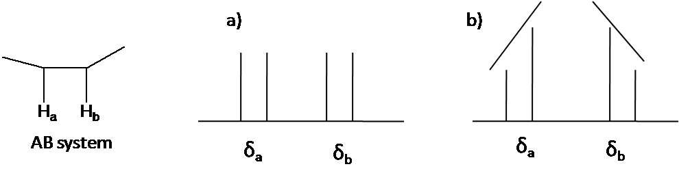
The 2nd order pattern is observed as leaning of a classical design: the inner peaks are taller and the outer peaks are shorter in instance of AB organization (Figure \(\PageIndex{4}\)). This is chosen the roof result.
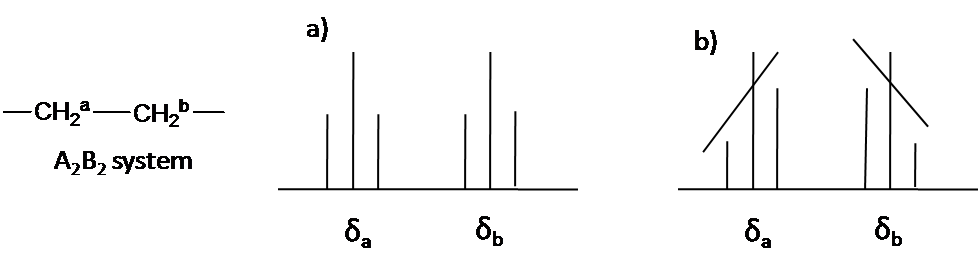
Here is other system as an instance: A2B2 (Figure \(\PageIndex{5}\)). The two triplet incline toward each other. Outer lines of the triplet are less than one in relative area and the inner lines are more than than 1. The middle lines have relative area 2.
Coupling constant (J Value)
Coupling constant is the strength of the spin-spin splitting interaction and the distance between the split lines. The value of distance is equal or different depending on the coupled nuclei. The coupling constants reflect the bonding environments of the coupled nuclei. Coupling abiding is classified by the number of bonds:
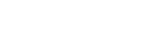
Geminal proton-proton coupling (2JHH)
Germinal coupling generates through two bonds (Figure \(\PageIndex{6}\)). 2 proton having geminal coupling are not chemically equivalent. This coupling ranges from -20 to 40 Hz. 2JHHdepends on hybridization of carbon cantlet and the bond angle and the substituent such as electronegative atoms. When South-grapheme is increased, Geminal coupling constant is increased: 2Jsp1>2Jsp2>2Jsp3 The bond angle(HCH) gives ascension to change 2JHH value and depend on the strain of the ring in the cyclic systems. Geminal coupling constant determines ring size. When bond angle is decreased, ring size is decreased so that geminal coupling constant is more than positive. If a atom is replace to an electronegative cantlet, Geminal coupling abiding move to positive value.
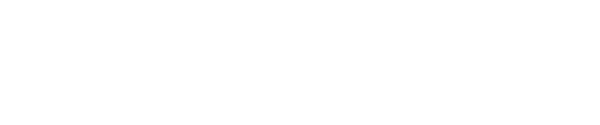
Vicinal proton-proton coupling (iiiJHH)
Vicinal coupling occurs though three bonds (Effigy \(\PageIndex{seven}\).). The Vicinal coupling is the most useful data of dihedral bending, leading to stereochemistry and conformation of molecules. Vicinal coupling abiding always has the positive value and is affected by the dihedral bending (?;HCCH), the valence bending (?; HCC), the bond length of carbon-carbon, and the furnishings of electronegative atoms. Vicinal coupling abiding depending on the dihedral angle (Figure \(\PageIndex{eight}\)) is given by the Karplus equation.
\[^3 J=7.0-0.5 \cos \phi+4.5 \cos ^{2} \phi\]
When ? is the 90o, vicinal coupling constant is zero. In addition, vicinal coupling abiding ranges from 8 to 10 Hz at the and ?=180o, where ?=0o and ?=180o means that the coupled protons have cis and trans configuration, respectively.
The valence angle(?;Effigy \(\PageIndex{viii}\)) too causes change of 3JHH value. Valence angle is related with band size. Typically, when the valence angle decreases, the coupling constant reduces. The distance between the carbons atoms gives influences to vicinal coupling constant
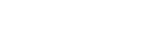
The coupling constant increases with the decrease of bail length. Electronegative atoms affect vicinal coupling constants then that electronegative atoms decrease the vicinal coupling constants.
Integral
Integral is referred to integrated peak surface area of 1H signals. The intensity is directly proportionally to the number of hydrogen.
13C NMR
Chemic Shift
Spin-Spin splitting
Comparing the aneH NMR, there is a big difference thing in the 13C NMR. The 13C- 13 C spin-spin splitting rarely get out betwixt side by side carbons because 13C is naturally lower arable (ane.1%)
- xiiiC-iH Spin coupling: thirteenC-oneH Spin coupling provides useful information about the number of protons attached a carbon atom. In example of 1 bond coupling (oneJCH), -CH, -CHtwo, and CH3 have respectively doublet, triplet, quartets for the thirteenC resonances in the spectrum. All the same, thirteenC-iH Spin coupling has an disadvantage for xiiiC spectrum interpretation. xiiiC-1H Spin coupling is hard to analyze and reveal construction due to a forest of overlapping peaks that consequence from 100% abundance of iH.
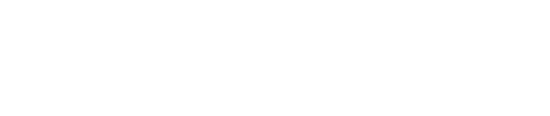
- Decoupling: Decoupling is the procedure of removing 13C-1H coupling interaction to simplify a spectrum and identify which pair of nuclei is involved in the J coupling. The decoupling 13C spectra shows merely one summit(singlet) for each unique carbon in the molecule(Figure \(\PageIndex{ten}\).). Decoupling is performed by irradiating at the frequency of 1 proton with continuous low-power RF.
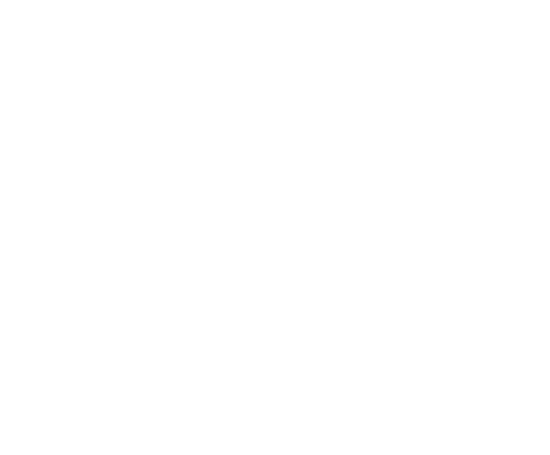
- Distortionless enhancement by polarization transfer (DEPT): DEPT is used for distinguishing between a CHthree group, a CH2 group, and a CH grouping. The proton pulse is set at 45o, xco, or 135o in the three separate experiments. The different pulses depend on the number of protons attached to a carbon atom. Figure \(\PageIndex{11}\). is an instance nigh DEPT spectrum.
2-dimensional NMR spectroscopy (COSY)
COSY stands for COrrelation SpectroscopY. COSY spectrum is more useful information about what is being correlated.
1H-1H COSY (COrrelation SpectroscopY)
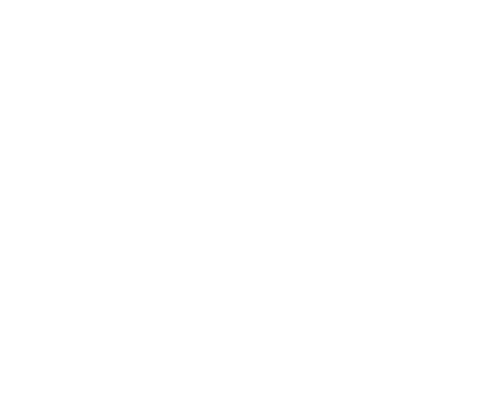
1H-1H COSY is used for clearly bespeak correlation with coupled protons. A point of entry into a COSY spectrum is ane of the keys to predict information from it successfully. Relation of Coupling protons is determined past cantankerous peaks(correlation peaks) and in the COSY spectrum. In other words, Diagonal peaks past lines ar east coupled to each other. Figure \(\PageIndex{12}\) indicates that there are correlation peaks between proton H1 and Htwo equally well as between H2 and Hiv. This means the Hii coupled to H1 and H4.
1H-13C COSY (HETCOR)
1H-13C COSY is the heteronuclear correlation spectroscopy. The HETCOR spectrum is correlated 13C nuclei with direct attached protons. 1H-13C coupling is 1 bond. The cantankerous peaks mean correlation between a proton and a carbon (Figure \(\PageIndex{13}\)). If a line does not have cross acme, this ways that this carbon atoms has no attached proton (e.thousand. a 4th carbon atom)

References
- Balc*, M., Basic p1 sH- and p13 sC-NMR spectroscopy. 1st ed.; Elsevier: Amsterdam ; Boston, 2005; p xii, 427.
- Breitmaier, Due east., Structure elucidation past NMR in organic chemical science : a practical guide. third rev. ed.; Wiley: Chichester, West Sussex, England, 2002; p xii, 258.
- Jacobsen, N. E., NMR spectroscopy explained : simplified theory, applications and examples for organic chemistry and structural biology. Wiley-Interscience: Hoboken, N.J., 2007; p fifteen, 668.
- Silverstein, R. K.; Webster, F. X., Spectrometric identification of organic compounds. sixth ed.; Wiley: New York, 1998; p 14, 482.
Exterior Links
- NMRShiftDB: a Free web database for NMR information : nmrshiftdb.chemie.uni-mainz.de/nmrshiftdb
- NMR database from ACD/LAbs : world wide web.acdlabs.com/products/spec_lab/exp_spectra/spec_libraries/aldrich.html
- NMR database from John Crerar Library : http://crerar.typepad.com/crerar_lib...h_ir_nmr_.html
Bug
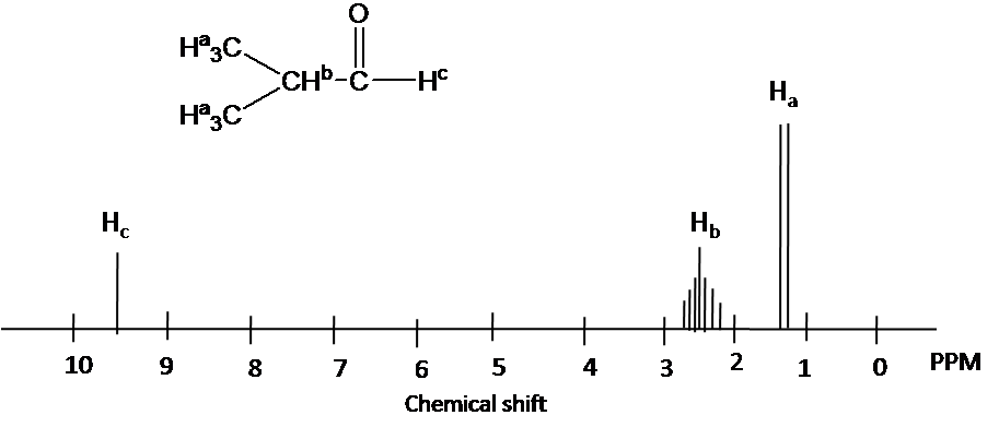
Draw the 1H NMR spectrum for ii-Hydroxypropane in CDCl3. Assume sufficient resolution to provide a first-society spectrum and ignore vicinal proton-proton coupling(3JHH)
Solution
1) the structure of two-hydoroxyporpane is drawn

Figure out which protons are chemically equivalent, i.due east., two methyl (-CH3) groups are chemic equivalent.
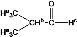
4) Splitting blueprint is determined by (N+1) rule: Ha is split up into two peaks by Hb(#of proton=1). Hb has the septet pattern by Ha (#of proton=vi). Hc has one pinnacle.(Note that Hc has doublet pattern by Hb due to vicinal proton-proton coupling.)
Contributors and Attributions
- Y'all Jin Seo

Source: https://chem.libretexts.org/Bookshelves/Physical_and_Theoretical_Chemistry_Textbook_Maps/Supplemental_Modules_%28Physical_and_Theoretical_Chemistry%29/Spectroscopy/Magnetic_Resonance_Spectroscopies/Nuclear_Magnetic_Resonance/NMR:_Experimental/NMR_-_Interpretation
Posted by: linseymarban.blogspot.com
0 Response to "How To Draw Nmr Spectra From Structure"
Post a Comment